Dr. Donald O. Freytes, Ph.D.; Joint Department of Biomedical Engineering at UNC-CH and NCSU

Category: Uncategorized

Latest News June 2023

We have had some great news to share from the REdesign Lab and its alums!  It took a while to update, but here is some recent news.

  1. Ms. Daphne Sze successfully defended her Master’s Thesis.
  2. Dr. Lewis Gaffney successfully defended his thesis and joined us for a while as Postdoc.  Dr. Gaffney is now an R&D scientist at BioAesthetics.
  3. Dr. Andreea Biehl joined as a postdoc and recently published a new paper on a novel testing platform for vocal fold tissue engineering.
  4. Dr. Ana Gracioso Martins published her work in the Journal of Controlled Release.

We are very proud of their accomplishments.

Congrats to Dr. Andreea Biehl!!

Congratulations to our newest graduate, Dr. Andreea Biehl!! She did an excellent job presenting her thesis titled, ” Platforms for Vocal Fold Tissue Engineering”. Special thanks to her committee, Matthew Fisher, Michael Daniele, Bill Polachek and Ashely Brown.


Congrats again Dr. Biehl!


New Paper Accepted at Tissue Engineering Part A

We are pleased to announce another paper acceptance. Amber Detwiler lead the study titled “Donor Age and Time in Culture Affect Dermal Fibroblast Contraction in an In Vitro Hydrogel Model” Amber started in our lab as a SIRI student working collaboratively with Kathryn Polkoff from Jorge Piedrahita’s Lab and Lewis Gaffney from our lab. Congrats to all of the author’s especially Amber D. for publishing her first paper as an undergraduate researcher!

Little is known about the impact of donor cell age and time in culture on the contraction of cellular, hydrogel-based skin grafts. These results show how cellular phenotypes of porcine fibroblasts differ based on donor age and time in culture. This information is beneficial when addressing important inconsistencies in biomanufacturing of bioengineered skin grafts and in vitro models. These findings are relevant to research and therapies using bioengineered skin graft models and the results can be used to increase reproducibility and consistency during the production of bioengineered skin constructs. The information from this study can be extrapolated to future in vivo studies using human dermal fibroblasts in an in vivo model to help determine the best donor age and time in culture for optimal wound healing outcomes or more reproducible in vitrotesting constructs.

Citation: Amber Detwiler, Kathryn Polkoff, Lewis Gaffney, Donald O. Freytes, and Jorge A. Piedrahita.Donor Age and Time in Culture Affect Dermal Fibroblast Contraction in an In Vitro Hydrogel Model.Tissue Engineering Part A.ahead of printhttp://doi.org/10.1089/ten.tea.2021.0217


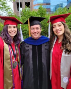
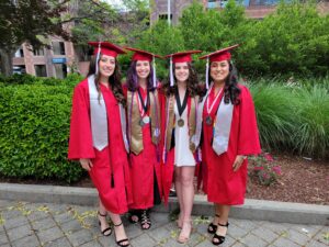
Congratulations to our 2022 Graduates!!

The ReDesign lab is happy to announce we had several graduates from the B.S. BME program as well as a graduate from the Ph.D. Program!

Lewis Gaffney finished his Ph.D. as a co-advised student by Matthew Fisher and Donald Freytes. He’s staying around with our lab as a new Post-Doc, where he’ll continue some studies with his Muscle-Tendon platform, as well as continuing work with ECM manufacturing as well as developing micro-tissue models for other physiological system. Congratulations Lewis!

Amber, Lewis and Caroline

Our wonderful undergraduates were: Amber Detwiler, Caroline Dau, Samantha Knapp, Bhairavy Puviindran. Amber Detwiler will be starting her graduate career at Duke University with Dr. Nenad Bursac. Caroline Dau will be working with a local company BioMilq, as an associate Engineer. Bhairavy is joining the Frank lab as a Post-Bac IRTA Fellow.  Samantha will be starting her career as a Research Technician at Anello.

Caroline, Amber and Samantha with another graduate

Congratulations to all of our new graduates, we are so happy you were a part of our lab!

Congratulations to Dr. Gaffney!!

We’re happy to announce the Lewis Gaffney successfully defended his thesis today! His work was titled “Crucial Structures of the Myotendinous Junction and Implications for Regenerative Medicine.”

Thanks to his Committee Members, Matthew Fisher (Co-Chair), Lauren Schnabel, Brian Diekmann and Jacque Cole!

Congratulations Lewis!


New Paper Accepted at Tissue Engineering Part A

We are excited to announce that Lewis Gaffney’s Paper “Extracellular Matrix Hydrogels Promote Expression of Muscle-Tendon Junction Proteins.” This paper was published in collaboration with Matt Fisher’s lab.

In this study we aimed to understand how muscle and tendon specific extracellular matrix hydrogels could promote muscle-tendon junction like proteins in muscle and tendon cells. Using a unique model to study the multi-tissue components, we broke down how specific micro-environments could affect individual cell types at the junction. Overall, there was increased protein expression of Paxillin and Type XXII Collagen (both specific MTJ proteins) when muscle and tendon cells were cultured in the tissue specific ECM hydrogels compared to Type I Collagen Hydrogels.


Congrats to all of the Authors!

Citation: Lewis S. Gaffney, Zachary G. Davis, Camilo Mora-Navarro, Matthew B. Fisher, and Donald O. Freytes.Extracellular Matrix Hydrogels Promote Expression of Muscle-Tendon Junction Proteins.Tissue Engineering Part A.Mar 2022.270-282.http://doi.org/10.1089/ten.tea.2021.0070

https://www.liebertpub.com/doi/full/10.1089/ten.tea.2021.0070

FIG. 1.

FIG. 1. ECM interactions and paxillin expression at the MTJ. (A) Graphical description of ECM–cell interactions at the MTJ. Where muscle and tendon meet, cells within one tissue interact with ECM of the other tissue. This interaction could be responsible for the production of MTJ-specific proteins such as paxillin, which is part of the integrin-mediated complex attaching muscle cells to tECM. (B) A mouse muscle tendon unit (Achilles tendon and gastrocnemius muscle) dissected from mouse hindlimb was sectioned for (C) hematoxylin and eosin staining, the dashed line indicating the junction of muscle and tendon, (D) anti-paxillin immunohistochemical staining. Arrows in (C) and in (D) indicate the junction of tendon (bottom) and muscle (top), scale bars are 50 μm. In (D), at the junction there is localization of paxillin expression in the ends of large muscle fibers, where the sarcolemma interacts and binds to tECM through the accumulation of focal adhesion complexes containing paxillin. MTJ, myotendinous junction; tECM, tendon extracellular matrix. Color images are available online.
© 2024 REdesign Lab

Theme by Anders NorenUp ↑